Nesara Agri
AboutHomeDiseases
Symptoms and Signs
Symptoms are the manifestation of disease expressed as a result of pathogen activity in the host plant. In some cases, pathogens produce structures like mycelium, spores, sclerotia, pycnidia, etc., on the surface of the infected parts. These are called ‘Signs’, e.g., mildews, rusts, etc. There are mainly three types of morpho- logical symptoms. Necrosis – Killing of plant tissues. Hypertrophy and hyperplasia – Over- development. Hypotrophy and hypoplasia – Under development and stunting.
Necrosis
Necrosis means death of cells, tissues and organs due to pathogen activity. Most destructive type and damage is irreversible.Almost all types of pathogens (fungi, bacteria, viruses, etc.) cause necrosis. Following are different kind of necrotic symptoms.
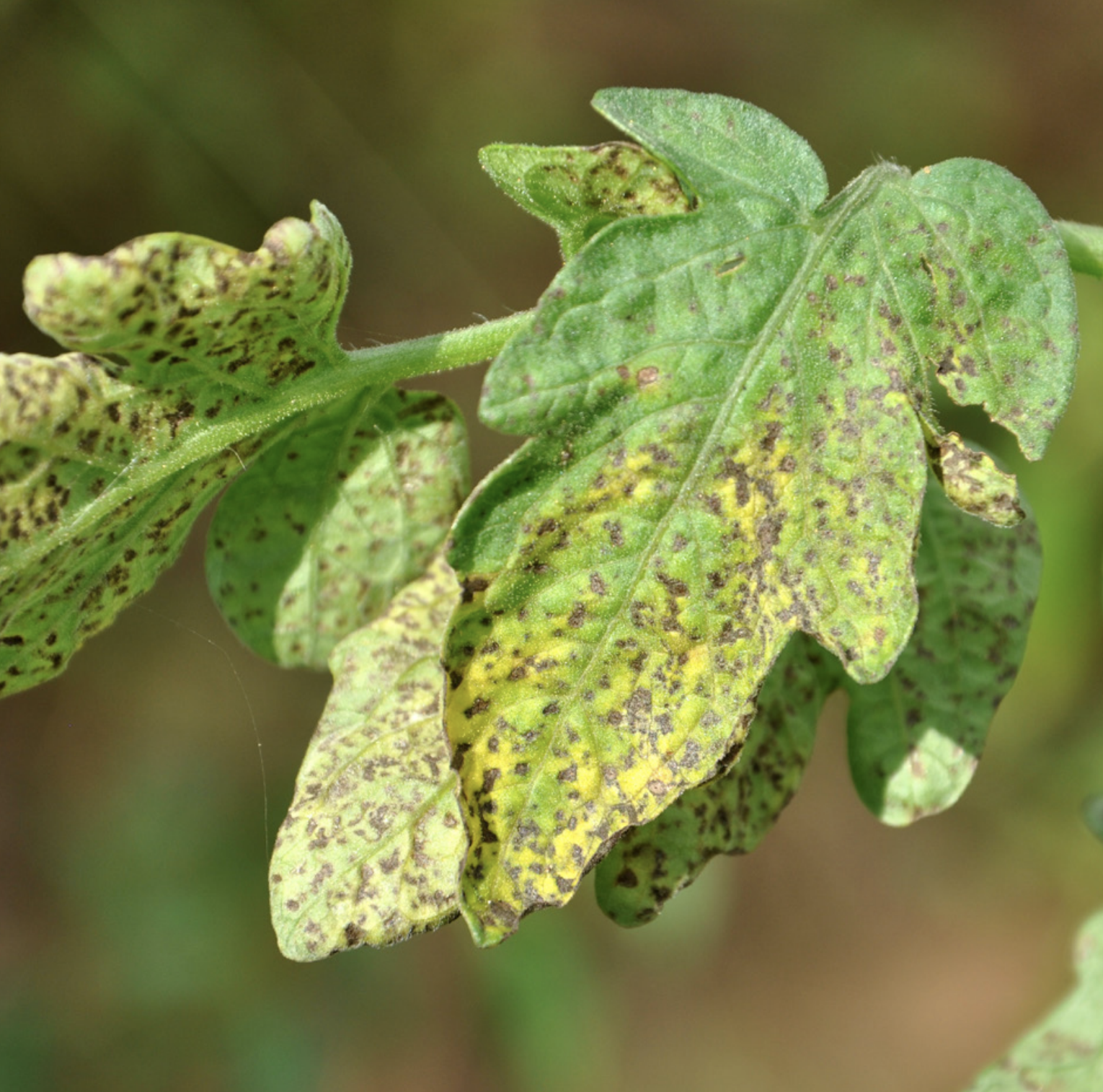
Spots
The most common necrotic symptoms that occur on leaves, floral parts and fruits.
They are well-defined localized areas of usually brown or tan necrotic tissue.
These spots may be surrounded by margins of purple or some other dark color.
Tiny spots are called ‘flecks’ or ‘specks’.
When dark threads of a fungal pathogen are seen on the spots, it is called ‘blotch’.
Sometimes fungal spots are raised and then they are called ‘tar spots’.
‘Shot hole’ spots are those where the dead and collapsed tissue shrink and fall off leaving a hole inside a spot.
In bacterial leaf spots they appear brown or water-soaked and usually remain localized
Blight and Blast
Initially, browning of infected plant parts occurs and then extensive death happens in a rapid manner.
The dead tissues soon disintegrate and give burnt or scorched appearances.
Scorch
Scorch resembles blight, but the scorch occurs in irregular patterns usually along the margins of leaves, and, sometimes between the veins.
Scald
Scald is the blanching of epidermal and adjacent tissues of fruit and, occasionally, of leaves.
Canker
A ‘Canker’ is really a symptom of an injury often associated with an open wound that has become infected by a fungal or bacterial pathogen.
Canker diseases frequently kill branches or structurally weaken a plant until the infected area breaks free, often during strong winds.
Citrus canker caused by the bacterium Xanthomonas axonopodis is one of the more common diseases.
Infection causes lesions on the leaves, stems and fruit of citrus trees, including lime, oranges, and grapefruit.
Die-back
Severe necrosis of twigs or branches happens from the tip and advances downwards Citrus canker caused by the bacterium Xanthomonas axonopodis is one of the more common diseases. towards the base is called ‘die-back’ or ‘stag head’.
The symptom often becomes apparent during the dormant season or shortly thereafter.
Species of Colletotrichum (Chillies), Botryodiplodia (Mango), Phomopsis (Neem) and other fungi cause this symptom.
Anthracnose
This fungal disease affects many plants, including vegetables, fruits and trees.
It causes dark, sunken lesions on leaves, stems, flowers and fruits.
It also attacks developing shoots and expanding leaves.
It can spread very quickly during rainy seasons.
Anthracnose is a general term for a variety of diseases that affect plants in similar ways.
On leaves, anthracnose generally appears first as small, irregular yellow or brown spots.
These spots darken as they age and may also expand, covering the leaves.
On vegetables, it can affect any part of the plant.
On fruits, it produces small, dark, sunken spots, which may spread.
In moist weather, pinkish spore masses form in the center of these spots.
Eventually, the fruits will rot.
On trees, it can kill the tips of young twigs.
It also attacks the young leaves, which develop brown spots and patches.
It can also cause defoliation of the tree.
Streak and Stripe
Streak represents elongated necrotic area along stems and leaf veins.
Stripe is also elongated necrotic area but confined to leaf tissues between the veins, particularly in monocots.
Ring spots
Ring spots are the common type of symptoms produced by virus pathogens.
They are characterized by the appearance of sometimes chlorotic but mostly necrotic rings on the leaves or also on the fruits and stems.
The ring spots may be present singly or in groups developing concentrically on infected parts.
Papaya ring spot, Tomato spotted wilt, Tobacco ring spot, etc., are common examples.
Rots
Rots represent dead and decomposed tissue on various parts of the host plant as a result of maceration and disintegration caused under the influence of various enzymes secreted by the pathogen.
Though the rotting may result due to nonparasitic causal agencies, the great majority of rots are caused by fungal or bacterial pathogens.
All parts are prone to rots and they are
- Root rot / Rhizome rot – in Ginger, Turmeric, Chrysanthemum, etc.
- Stem rot of Rice caused by Leptosphaeria salvini.
- Bud rot – Coconut, Arecanut caused by Phytophthora palmivora.
- Fruit rot - Most of the vegetables have fungal fruit rots.
- Ear and seed rot- Various fungi cause ear rots in Maize.
- Leaf rot - Black rot of crucifers (Xanthomonas campestris pv. campestris)
- Tuber rot – Brown rot of Potato (Ralstonia solanacearum).
Scabs
Scabs are rough, crust-like localized lesions usually slightly raised or sunken and cracked giving a scabby appearance.
Though they can occur on every part of the host but are frequently evident on fruits and tubers
Wilts
Wilt is usually a generalized secondary symptom where leaves and shoots lose their turgidity, become flaccid and finally droop (wilt).
Normally this happens due to blocking of xylem vessels by the pathogen itself or by substances secreted by it.
Wilts may be sudden without yellowing (bacterial wilt of solanaceous crops) or gradual, partial and slow wilt with yellowing of leaves and finally total wilt as in the case of Fusarium and Verticillium wilts.
Plants also wilt because of rotting of roots and stems.
Rust pustules
These are the characteristic symptoms of rust fungi.
All rust fungi are obligate pathogens.
Rust pustules are normally erumpent; submerged, small or big in size.
Generally rust pustules are surrounded by chlorotic halos.
Though they are common on leaves, they can be found on stems also.
Normal colour of the pustules is red or yellow, however, brown to dark colors are also not uncommon in fungi belonging to Basidiomycetes.
Pustules may be of whitish colour in white rusts caused by Albugo candida under Oomycetes.
And may be red in red rust caused by Cephaleurus parasitica, a green alga
Bleeding and Exudations
Refers to the abnormal secretion or oozing out of watery or slimy products from the affected plant parts, which may or may not set into hard masses.
It is the major symptom of some fungal diseases.
Citrus gummosis, Gummy stem blight in Cucurbits and stem bleeding in Coconut and Arecanut are some of the examples.
Damping-off
Sudden collapse of seedlings specially in nursery bed or tray.
Fall over of the seedlings due to weakening of the base of the stem.
Species of Pythium and Rhizoctonia are the common fungi that cause this.
Excess moisture or dampness causes this disease.
Mildews
Mildews represent the chlorotic or necrotic areas generally covered with sporulation of the fungal pathogen.
There are two types of mildews namely ‘Downy’ and ‘Powdery’.
Downey Mildews
Downy mildews are caused by a group of Oomycetous ‘fungal’ pathogens belonging to family Peronosporaceae.
Downy mildews occur on both monocots and dicots.
Upper surface of the leaf show chlorotic spots (pale yellow/purplish / yellowish, etc.) of varying size and form which later merge into surrounding green tissues.
The corresponding lower surface is covered with white cottony or downy growth, hence the name ‘Downy Mildew’.
However, under severe disease condition, the cottony growth and sporulation can happen on upper surface also.
In some cases the infection will occur on floral parts and even on fruits.
In cereal Downy Mildews, phyllody of the inflorescence (green ear) is also caused.
Powdery Mildews
Powdery mildews are caused by fungi belonging to Ascomycetes and family Erysiphaceae.
These are also obligate pathogens like Rust fungi.
Powdery mildew is the most common disease occuring on a very large range of crops.
The symptoms are typical and very easy to recognize owing to its powdery appearance.
They produce chlorotic or necrotic areas covered superficially growing mycelia and fructifications of the pathogen.
Black minute fruiting bodies are also visible at the end of the season.
Hyperplasia and Hypertrophy (Over Development)
Many pathogens, through their biochemical activity induce hormonal imbalance.
This results in excessive growth of host tissues and causes abnormal increase in size of affected organs.
It is brought about by one or both of the two processes known as hyperplasia and hypertrophy.
Hyperplasia is the abnormal increase in the size of plant organs due to increase in number of cells by increased cell division rate.
In hypertrophy the increased size of the organ is due to increase in the size of cells of a particular tissue.
Galls
Galls are localized enlargements on various plant parts in the form of small pustules or warts, larger tubercles, tumours or masses of cells making a morbid outgrowth of either fleshy or woody texture, in which host tissues and pathogens unite.
Knots may be fleshy or woody in their texture.
Nematode like Meloidogyne incognita, fungi like Protomyces macrosporus, bacteria like Agrobacterium tumefaciens are some of the common examples.
Warts
Warts are protuberances developed on tubers and stems generally giving warty or discolored Cauliflower-like appearance.
Good example is wart disease of Potato caused by Synchytrium endobioticum.
Leaf Curl
These symptoms represent irregular and extensive wrinkling and furrowing of leaves due to reduced growth of veins in comparison with the growth of laminar tissue resulting in sunken veins and raised up laminar tissue leading to the curling of the leaves.
Papaya, Tomato, Cotton, Tobacco, Chilli crops are most susceptible to leaf curl by whitefly transmitted viruses.
Hypotrophy and Hypoplasia
In some diseases, infected cells and tissues slowdown in development resulting in the infected plant parts thereof being arrested in their growth as a result of degeneration of the cells (hypotrophy) or subnormal cell divisions (hypoplasia).
Though some of the fungal and bacterial diseases exhibit these symptoms, viruses produce more of these symptoms.
Chlorosis
Destruction of or inhibition of chlorophyll formation is called chlorosis.
The leaves with less chlorophyll appear pale green or yellowish in color.
Extreme case of chlorosis is ‘blanching’ or ‘bleaching’
Chlorosis is commonly caused by viruses.
However, certain nutrient deficiencies also cause chlorosis.
Variety of chlorotic symptoms are caused by viruses viz., mosaic, mottle, vein-chlorosis, vein-clearing, vein-banding, etc.
Mosaicing
Irregular intermingling of normal green with light green, yellow or white chlorotic areas imparting a mosaic-like pattern.
Tomato mosaic, Tobacco mosaic, Pumpkin mosaic, Yellow vein mosaic of Okra, etc.
Leaf narrowing
The infected leaves generally become narrow due to reduced growth of laminar tissue, but the veins and midrib remain normal in growth.
In extreme cases only the main veins remain normal while laminar tissue is almost completely absent, e.g. rat tail symptom in Tomato (CMV), Papaya (PRSV), etc.
Stunting or dwarfing
Refers to the reduction in size of either the whole plant or a specific plant part.
It results in reduced size and loss of vigour.
Stunting may be caused by viral, bacterial, fungal or nematode infections.
Insect or mite feeding can also cause stuntingor dwarfing.
Even abiotic factors like lack of water, imbalance of soil nutrients, excess light, chemical or mechanical injuries can cause stunting.
Stunting due to abiotic factors can easily be corrected.
Rosetting
Rosetting means shortening of internodes of the infected plant along with the clumping of the foliage together at the apices of branches.
Fasciation
Fasciation also known as cresting, is a relatively rare condition of abnormal growth in vascular plants.
Here the apical meristem (growing tip), which normally is concentrated around a single point and produces approximately cylindrical tissue, instead becomes elongated perpendicularly to the direction of growth, thus, producing flattened, ribbon-like, crested (cristate), or elaborately contorted tissue.
Fasciation may also cause plant parts to increase in weight and volume in some instances.
The phenomenon may occur in the stem, root, fruit or flower head.
Some plants are grown and prized aesthetically for their development of fasciation.
Any occurrence of fasciation has several possible causes, including hormonal, genetic, bacterial, fungal, viral and environmental causes.
Transformation or Replacement of Plant Organs
Normal plant organs particularly floral organs are replaced by a structure of the pathogen, causing severe yield losses.
In case of smut, the ovary is transformed into structure containing smut spores.
In ergot disease, the ovary is invaded by the fungus and transforms it into scelotial body.
In Udbatta disease of Rice, which is also called false ergot, the seeds become pseudosclerotia.
Little leaf disease
This is a serious mycoplasma disease of Brinjal.
The disease is transmitted by leaf hoppers (Cestius (Hishimonus) phycitis and Amrasca biguttula).
The leaves of the infected plants in the early stages are light yellow in colour, reduced in size and are malformed.
Disease affected plants are generally shorter in stature bearing a large number of branches, and leaves much much smaller than healthy plants.
The petioles get shorter considerably, many buds appear in the axil of leaves and internodes get shortened thus giving the plants a bushy appearance (rosetting/ witches broom symptoms).
Flower parts are deformed leading the plants to be sterile.
Infected plants do not bear any fruit.
However, if any fruit is formed it becomes hard and tough and fails to mature.
Recently this disease is also affecting Beans crop
Smuts
Smuts refer to sooty or charcoal-like powdery mass usually appearing on floral organs, particularly the ovary.
However, they can occur on leaf, stem and roots.
They are caused by Basidiomycetes belonging to order Ustilaginales.
Smut infected tissues are either destroyed or replaced by black smut spores or they are first stimulated to divide and enlarge to produce a swelling or gall of varying size and then destroyed and replaced by smut spores.
Corn, Sorghum, Wheat and Rice are the common smut hosts.
Ergots
The ergot causing fungus infects the florets and develops in the ovaries, producing initially copious creamy, pink, or red colored sweet sticky liquid called honeydew.
The honeydew can drip down onto the upper leaves making them sticky.
Often pollen and anther sacs adhere to the honeydew.
Subsequently, long dark colored hard structures, sclerotia, develop from infected florets, first dark at the tip and then completely black.
Udbatta disease or incense rod disease is also called ‘False Ergot of Rice’ as it is not Ergot in real sense.
Transformation of floral organs
Floral organs are transformed into leafy structures.
Phyllody refers to the development of floral parts into leafy structures, generally caused by virus or Phytoplasma infection such as aster yellows.
However, in case of Downy Mildew of Finger millet, Pearl millet and Maize (male tassel) the inflorescence can show phyllody.
Phyllody causes the affected plant to become partially or entirely sterile.